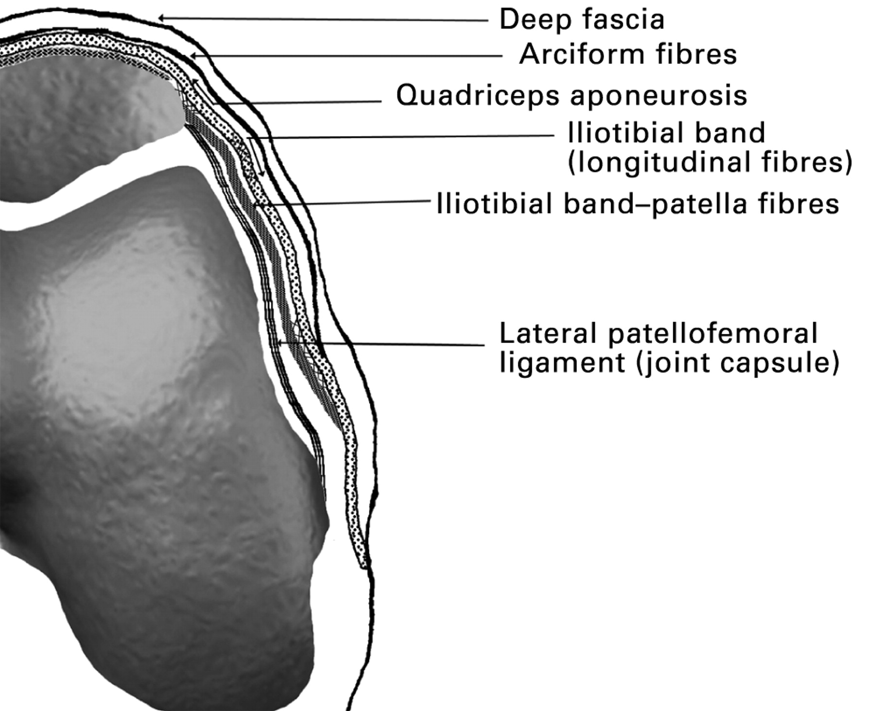
Definition and Anatomy

The medial retinaculum is a fibrous band of connective tissue that runs along the medial (inner) aspect of the ankle joint. It is a thickened portion of the deep fascia of the leg and serves to hold the tendons of the tibialis posterior and flexor digitorum longus muscles in place as they pass behind the medial malleolus.
The medial retinaculum is a relatively thin but strong structure. It originates from the medial malleolus and inserts onto the navicular bone. It is continuous with the plantar fascia and the flexor retinaculum.
The medial retinaculum forms the roof of the tarsal tunnel, a narrow passageway through which the tendons of the tibialis posterior, flexor digitorum longus, and flexor hallucis longus muscles pass. The tarsal tunnel is located on the medial side of the ankle and is bounded by the medial malleolus, the calcaneus, and the navicular bone.
Function and Biomechanics

The medial retinaculum plays a vital role in maintaining the stability and functionality of the wrist joint. It acts as a strong, fibrous band that stabilizes the tendons passing through the carpal tunnel, preventing them from dislocating or bowstringing during wrist movements.
Contribution to Wrist Stability
- Prevents Tendon Bowstringing: The medial retinaculum prevents the tendons from bulging or “bowstringing” out of the carpal tunnel during wrist flexion. This is crucial for maintaining the tendons’ alignment and preventing friction or irritation against the surrounding structures.
- Resists Wrist Extension: The medial retinaculum acts as a strong barrier against excessive wrist extension, which could potentially lead to instability or dislocation of the carpal bones.
Biomechanical Properties, Medial retinaculum
The medial retinaculum is composed of dense, collagenous fibers that provide tensile strength and flexibility. Its biomechanical properties contribute to its ability to withstand the forces generated during wrist movements:
- High Tensile Strength: The collagen fibers within the retinaculum provide exceptional resistance to stretching forces, allowing it to maintain its integrity under significant loads.
- Elasticity: The retinaculum exhibits a degree of elasticity, enabling it to stretch and recoil during wrist movements without losing its structural integrity.
- Low Friction: The inner surface of the retinaculum is smooth and lined with synovial fluid, reducing friction between the tendons and the surrounding structures, facilitating smooth gliding during wrist flexion and extension.
Clinical Significance: Medial Retinaculum

The medial retinaculum plays a pivotal role in maintaining the stability of the wrist joint. Injuries or conditions affecting this structure can lead to pain, discomfort, and functional limitations.
Common Injuries and Conditions
- Carpal Tunnel Syndrome: A condition where the median nerve is compressed within the carpal tunnel, which is formed by the medial retinaculum and the carpal bones.
- De Quervain’s Tenosynovitis: Inflammation of the tendons that pass through the first dorsal compartment, located adjacent to the medial retinaculum.
- Medial Retinaculum Tear: A rupture of the medial retinaculum, often resulting from a forceful wrist flexion or pronation.
Symptoms and Diagnosis
Symptoms of medial retinaculum injuries may vary depending on the specific condition. However, common symptoms include:
- Pain and tenderness over the medial aspect of the wrist
- Numbness or tingling in the thumb, index, and middle fingers
- Difficulty grasping or pinching objects
Diagnosis involves a physical examination, medical history, and imaging tests such as X-rays or magnetic resonance imaging (MRI) to confirm the underlying condition.
Treatment Options
Treatment for medial retinaculum injuries depends on the severity and nature of the condition. Non-surgical options include:
- Rest and immobilization
- Corticosteroid injections
- Physical therapy to improve range of motion and strength
Surgical intervention may be necessary in cases where non-surgical treatments fail to provide relief. Surgical options include:
- Carpal Tunnel Release: A procedure to divide the medial retinaculum and release pressure on the median nerve.
- De Quervain’s Tenosynovitis Release: A procedure to open the first dorsal compartment and release pressure on the affected tendons.
- Medial Retinaculum Repair: A procedure to suture a torn medial retinaculum.
The medial retinaculum, a fibrous band that stabilizes the flexor tendons in the wrist, plays a crucial role in hand function. Its significance extends beyond the realm of anatomy, mirroring the pivotal role of Mark Cuban’s sale of the Mavericks in reshaping the NBA landscape.
Just as the medial retinaculum provides structural support, Cuban’s decision has sparked a ripple effect that will reverberate throughout the league.
The medial retinaculum, a band of connective tissue that holds the flexor tendons in place, plays a crucial role in hand function. Like the unwavering support of Bill Russell’s wife , Maureen Russell, who stood by his side throughout his illustrious basketball career, the medial retinaculum provides stability and strength to the tendons, allowing for precise and delicate movements of the fingers.
Like a guardian angel, the medial retinaculum stands watch over the tendons of the wrist, protecting them from harm. Its intricate dance with the wyc grousbeck reminds us of the delicate balance between strength and flexibility. As the tendons glide through the retinaculum’s embrace, they find solace and guidance, knowing that they are sheltered from the perils of everyday life.
The medial retinaculum, a fibrous band stabilizing the wrist joint, whispers secrets of resilience. Its tensile strength echoes the tenacity of the cowboys who once roamed the vast prairies. As the news of the cowboys trade CeeDee Lamb reverberates, it’s a poignant reminder of the unyielding spirit that binds the gridiron and the human body, where strength and flexibility dance in harmony.
The medial retinaculum, a fibrous band on the palmar surface of the wrist, is a vital structure for stabilizing the wrist joint. Its intricate interplay with tendons and ligaments ensures smooth hand movements. Speaking of precision and agility, did you know that tennis legend John McEnroe recently shared his insights on the latest trends in the sport?
John McEnroe’s latest news offers a glimpse into his astute observations on the evolution of tennis strategy. Returning to the medial retinaculum, its role in supporting the wrist’s range of motion is truly remarkable.